Synaptogenesis: How Neurons Form Connections and Reshape the Brain

Synaptogenesis spans development and adulthood, involving axon guidance, synaptic differentiation, pruning, and circuit remodeling.
Synaptogenesis is the process by which neurons form synaptic connections, supporting early neural development and continuing throughout life to contribute to learning, memory, and neuroplasticity. The article reviews its key stages: growth cones sense attractive and repulsive molecular cues to guide axons to their targets; upon contact, pre- and postsynaptic cells each assemble their own molecular machinery to complete bidirectional synaptic differentiation; during development, the nervous system overproduces synapses and then refines circuits through selective pruning; in the adult brain, synaptogenesis manifests more as remodeling of existing circuits in response to experience and neural activity. The article emphasizes that brain plasticity is supported by concrete cellular and molecular events, and understanding synaptogenesis is the foundation for grasping more complex brain functions.
Synaptogenesis is the process by which neurons form synaptic connections with one another and with other cells. It is both a fundamental step in nervous system development and a process that continues throughout life, contributing to learning, memory, and neuroplasticity. In other words, the brain isn't hardwired once development is complete—it continuously adjusts its connections, driven by experience and neural activity.
How Axons Find the Right Target
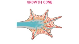
During early neural development, growing axons extend toward other cells. At the tip of each growing axon is a specialized structure—the growth cone—that steers the axon toward its target.
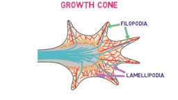
The growth cone extends two types of structures: finger-like filopodia and sheet-like lamellipodia. These continuously "sample" molecular guidance cues in the surrounding environment. Some cues attract the growth cone, while others repel it; together they steer the axon along a specific path. This balance of attraction and repulsion is one of the key mechanisms enabling precise wiring of neural circuits.
The growth cone is a highly dynamic, specialized structure at the axon tip, driven in its movement by the actin cytoskeleton. Filopodia are slender, F-actin-rich protrusions that probe the environment through continuous extension and retraction; lamellipodia are broader sheet-like structures that provide forward thrust for the growth cone. The growth cone surface carries a variety of guidance receptors, such as DCC, Robo, and Eph, which recognize secreted or membrane-bound guidance molecules including Netrin, Slit, and Ephrin. The signaling pathways mediated by these receptors ultimately converge on the Rho GTPase family (RhoA, Rac1, Cdc42), which regulates cytoskeletal rearrangement and thereby translates chemical signals into turning behavior of the growth cone. Understanding this signal transduction chain is the molecular basis for understanding how axons navigate with precision.
After Contact: Synaptic Differentiation
When the growth cone reaches its target cell, molecular interactions between the surfaces of the two cells help establish contact and initiate synaptogenesis. This process is essentially synaptic differentiation: both cells begin developing the specialized structures required for synaptic communication.
The presynaptic cell forms synaptic vesicles and the proteins involved in neurotransmitter release; the postsynaptic cell forms neurotransmitter receptors and associated proteins, building a functional synapse. Because each side assembles its own distinct molecular machinery, synaptogenesis is not a one-sided "wiring" job but a bidirectional, collaborative construction.
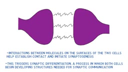
Synaptic differentiation relies on interactions between trans-synaptic adhesion molecules, such as the binding of neurexin to neuroligin and synaptic cell adhesion molecules (SynCAMs). These adhesion molecules not only physically connect the two cells but also recruit scaffold proteins (such as PSD-95 and SAP102) through their intracellular domains, thereby organizing the assembly of the postsynaptic density (PSD). In addition, synaptic differentiation involves local signal transduction: kinases such as protein kinase A on the presynaptic side and CaMKII on the postsynaptic side are activated, driving vesicle clustering and receptor anchoring. The temporal sequence and spatial localization of these molecular events determine whether a synapse can mature from initial contact into a functional connection.
Build More, Then Prune: The Significance of Synaptic Pruning
During development, the nervous system often produces more synapses than are ultimately needed. These excess connections are subsequently removed through synaptic pruning. Pruning refines neural circuits and improves the efficiency of communication between neurons.
This "overproduce first, then selectively retain" strategy allows the nervous system to keep valuable connections based on actual use while eliminating those that are used less often. Neuroscience popular science content from various sources is highly consistent in describing this mechanism: synaptic pruning is a core step in circuit refinement during development and an important entry point for understanding neuroplasticity.
The cellular and molecular mechanisms of synaptic pruning involve the active participation of glial cells. In the central nervous system, microglia tag weak or excess synapses through the complement system (such as C1q and C3) and engulf them via the complement receptor CR3; astrocytes participate in pruning through phagocytic receptors such as MEGF10 and MERTK. This process is activity-dependent: frequently used synapses tend to be retained, while low-activity synapses are more likely to be tagged and removed. Abnormal pruning is associated with various neurodevelopmental disorders—for example, hypotheses suggest that autism spectrum disorder may involve insufficient or excessive pruning. Thus, synaptic pruning is not only a step in circuit refinement but also an important window into understanding the mechanisms of brain disease.
Synaptogenesis in the Adult Brain
Synaptogenesis does not occur only during development. It also occurs in the adult brain, though in a different form: it manifests more as remodeling of existing neural circuits in response to experience and neural activity, rather than the axon growth and guidance seen in early development.
This distinction is worth noting. Developmental synaptogenesis focuses on building connections "from scratch," whereas adult synaptogenesis focuses on adjusting the weights and structure of existing networks. Together they support the enduring capacity for learning and memory, and explain why experience can change the way the brain is wired.
Structurally, adult synaptogenesis manifests primarily as dynamic changes in dendritic spines. Dendritic spines are tiny protrusions on the postsynaptic membrane of excitatory synapses, and their morphology (thin, mushroom-shaped, stubby) is closely related to synaptic strength. During learning and memory, new dendritic spines can form within minutes to hours, while existing spines can enlarge or shrink. This structural plasticity depends on NMDA receptor activation and actin remodeling, and is regulated by neurotrophic factors such as BDNF. Unlike during development, adult synaptogenesis typically does not involve long-distance axon guidance but rather local reorganization between existing axon terminals and dendrites. In addition, adult neurogenesis (mainly in the hippocampal dentate gyrus) can also generate new synaptic connections, though its contribution to the adult human brain remains debated.
The Value of Understanding Synaptogenesis
From growth cone guidance to synaptic differentiation, and from pruning to circuit remodeling in adulthood, synaptogenesis runs through the entire chain of nervous system construction and adjustment. It reminds us that brain plasticity is not an abstract concept but is supported by a series of concrete cellular and molecular events. For anyone interested in learning mechanisms, neural development, or brain disease research, grasping the basic processes of synaptogenesis is the foundation for understanding more complex brain functions.
One-line Summary
Synaptogenesis spans development and adulthood, involving axon guidance, synaptic differentiation, pruning, and circuit remodeling.
Paragraph Summary
Synaptogenesis is the process by which neurons form synaptic connections. It supports early neural development and continues throughout life, contributing to learning, memory, and neuroplasticity. The article systematically reviews its key stages: growth cones sense attractive and repulsive molecular cues through filopodia and lamellipodia, guiding axons to their targets; upon contact, presynaptic cells form synaptic vesicles and release-related proteins while postsynaptic cells form receptors and associated proteins, completing bidirectional synaptic differentiation; during development, the nervous system overproduces synapses and then selectively retains effective connections through synaptic pruning to refine circuits; in the adult brain, synaptogenesis manifests more as remodeling of existing circuits in response to experience and neural activity. The article emphasizes that brain plasticity is supported by concrete cellular and molecular events, and that understanding the basic processes of synaptogenesis is the foundation for grasping more complex brain functions.
Related articles

Gemini 4 Neon Leak: A Potential Strong Contender Against Opus 5.5
A Hacker News thread suggests Gemini 4 Neon looks like an Opus 5.5 competitor. This article unpacks the product positioning behind this community signal, shifts in the top-model competitive landscape, and what it means for developers' model selection strategy.

Claude Fable 5.2's Self-Authored Short Film 'Fable' Sparks Discussion
A short film 'Fable,' credited to Claude Fable 5.2 for script, visuals, and directing, appears on Hacker News. This article analyzes the significance of full-pipeline AI creation, community reactions, and what it means for content creators.

Anthropic IPO Pricing: A Signal of AI Giants Going Public
Anthropic's IPO pricing draws attention. This article analyzes its potential impact on AI industry valuation, commercialization paths, and the competitive landscape, and highlights public-market expectations and challenges for AI companies.