A Population-Supervised Framework for Inferring 3D Cell Biophysical Properties from 2D Microscopy Images

Population-supervised framework infers MCV/RDW/MCH from 2D red cell images with formal identifiability analysis.
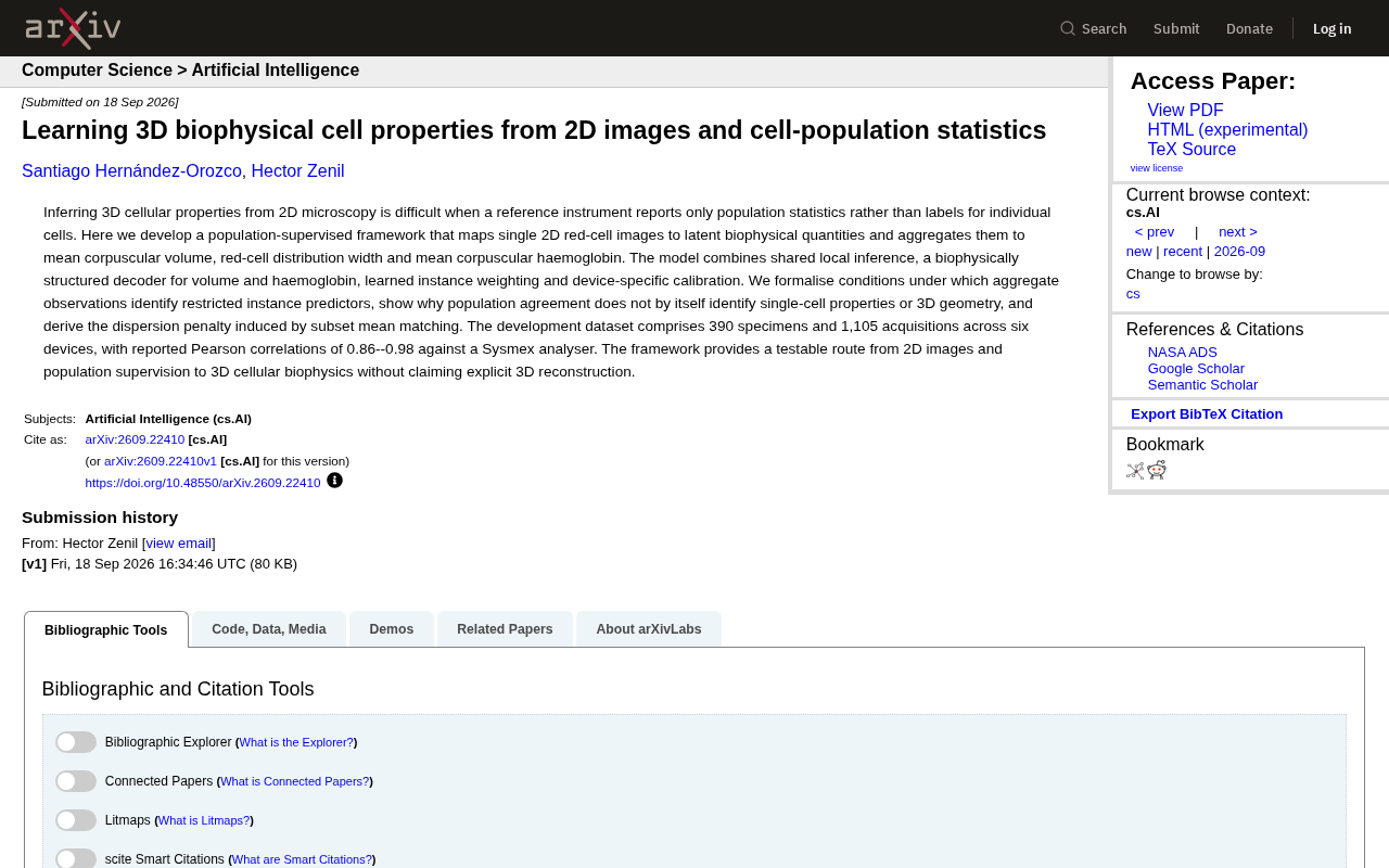
This study addresses the challenge of reference instruments providing only population statistics without single-cell labels, proposing a population-supervised framework that maps 2D red blood cell microscopy images to biophysical latent variables and aggregates them into clinical metrics (MCV, RDW, MCH). Built on a biophysically structured decoder, learned instance weighting, and device-specific calibration, the model achieves Pearson correlations of 0.86–0.98 against Sysmex analyzers across 1,105 acquisitions from 390 specimens on six devices. Notably, the authors formally analyze identifiability conditions and explicitly state that population consistency alone cannot verify accurate reconstruction of single-cell properties or 3D geometry.
Background: When Reference Instruments Only Provide Population Statistics, Not Single-Cell Labels
Inferring three-dimensional cell properties from two-dimensional microscopy images has long been a challenging problem in computational biology. The core tension is this: reference instruments commonly used in clinical and laboratory settings (such as Sysmex hematology analyzers) typically report only population-level statistics, not per-cell annotations. This means researchers lack the single-cell ground truth needed for supervised learning, making traditional supervised learning paradigms difficult to apply directly.
This arXiv study proposes a population-supervised framework that aims to work around the absence of single-cell labels by mapping individual 2D red blood cell images to underlying biophysical quantities, which are then aggregated into clinically relevant metrics.
Sysmex hematology analyzers are fully automated blood cell analysis devices widely used in clinical hematology. They operate on the principles of electrical impedance (the Coulter principle) and radiofrequency/laser light scattering, measuring changes in electrical signals as cells pass through a microaperture to count cells and characterize their volume distribution. The instrument outputs aggregate statistics across thousands to tens of thousands of cells in a blood sample — such as MCV (mean) and RDW (distribution width) — without labeling each cell individually. While this measurement approach is efficient and reliable in clinical practice, it means machine learning researchers cannot directly obtain instance-level supervision signals like "the volume of cell i = X fL." Bridging the gap from "single-cell predictions" to "population statistics" is precisely the core engineering and theoretical challenge this research addresses.
Methodological Framework: From Single-Cell Mapping to Population Aggregation
The framework's core logic is to first perform inference at the single-cell level, then aggregate up to population statistics, using population-level observations as the supervisory signal. The model consists of several key components:
Shared Local Inference and Biophysical Decoder
The model performs shared local inference on each 2D red blood cell image to extract underlying biophysical quantities. A key design choice is a biophysically structured decoder specifically designed to estimate cell volume and haemoglobin content. Embedding physical constraints into the decoder encourages the latent representations to correspond to biologically meaningful quantities rather than opaque black-box features.
Instance Weighting and Device Calibration
The framework also incorporates learned instance weighting and device-specific calibration. The former allows the model to assign different weights to individual cell samples, while the latter accounts for systematic differences across multiple devices — a critical consideration in real-world multi-device acquisition scenarios.
Single-cell estimates are ultimately aggregated into three clinical metrics: mean corpuscular volume (MCV), red cell distribution width (RDW), and mean corpuscular haemoglobin (MCH).
MCV, RDW, and MCH are core red blood cell parameters in the complete blood count (CBC), used clinically for anemia classification and differential diagnosis. MCV reflects the average red cell size (normal range ~80–100 fL); low values suggest microcytic anemia (e.g., iron deficiency), while high values suggest macrocytic anemia (e.g., folate/B12 deficiency). RDW measures the degree of variability in cell size, with elevated values indicating anisocytosis. MCH reflects the absolute amount of haemoglobin per red cell, jointly determining oxygen-carrying capacity along with cell volume and haemoglobin concentration. All three are population statistics, which aligns naturally with the aggregated predictions this framework produces — and provides the clinical rationale for choosing them as validation targets.
Theoretical Contribution: What Population Consistency Can and Cannot Identify
A methodological highlight of this work is that it goes beyond engineering implementation to provide a formal analysis of identifiability. The authors explicitly characterize the conditions under which aggregate observations can identify restricted instance predictors.
More importantly, the authors honestly acknowledge the limits of this paradigm: population-level consistency alone cannot identify single-cell properties or 3D geometric structure. In other words, even if a model's population statistics closely match those of the reference instrument, this cannot be taken as evidence that the model accurately reconstructs the true 3D morphology of individual cells. The study also derives the dispersion penalty introduced by subset mean matching, providing theoretical grounding for understanding model behavior.
This candor about the method's limitations is precisely what makes the research commendable — the authors explicitly state they do not claim explicit 3D reconstruction, but rather offer a verifiable pathway from 2D images and population supervision toward 3D cell biophysics.
Identifiability is a foundational concept in statistics and machine learning, referring to whether model parameters can be uniquely determined from observable data. In traditional supervised learning, per-sample labels generally ensure identifiability; but when supervisory signals come only from population aggregates (such as means and variances), the conditions for identifiability become far more restrictive. Intuitively, the same population statistics can be generated by infinitely many different single-cell distributions — the technical backdrop known as "aggregation irreversibility" or the "ecological fallacy." This study formally characterizes what constraints (e.g., restrictions on the parameter family) are sufficient for aggregate observations to uniquely determine restricted instance predictors. The significance of this analysis is twofold: it provides theoretical guarantees for model design, while honestly delineating the method's upper bound — for single-cell properties that fall outside these constraints, population supervision cannot in principle provide reliable inferential support.
Experimental Data and Validation Results
The development dataset includes 390 specimens across 1,105 acquisitions from six devices. In comparison with the Sysmex analyzer, the model achieved Pearson correlation coefficients of 0.86 to 0.98.
This correlation range is quite strong for a method that relies solely on population supervision without any single-cell annotations. It demonstrates that with appropriate structural design and aggregated supervision, the model can indeed capture biophysical information highly correlated with clinical hematology metrics. Validation across six devices also provides some evidence of the framework's cross-device generalization and calibration capabilities.
Significance and Outlook
The value of this research extends beyond hematology applications — it proposes a generalizable weakly supervised inference paradigm for situations where only population statistics are available and individual labels are absent, while maintaining theoretical rigor in inferring individual-level properties.
This approach to "inferring individual quantities from aggregate observations" has implications for medical imaging, single-cell omics, and broader scientific measurement contexts. At the same time, the authors' clear articulation of identifiability boundaries highlights a critical pitfall for future work: good population fit ≠ accurate individual inference. Future efforts that incorporate partial single-cell ground truth for semi-supervised validation, or leverage true 3D imaging data for verification, would further strengthen the credibility of this pathway.
Related articles

Chain of Thought Isn't Just for AI: How Humans Can Use CoT to Improve Thinking Quality
Chain of Thought isn't just a prompt trick for large models. Starting from a Hacker News discussion, this article analyzes why CoT works for human thinking too, breaks down actionable ways to transfer it, and highlights the key differences between human CoT and machine CoT.
Cloudflare Teams Up with OpenAI: Closi…
Cloudflare Teams Up with OpenAI: Closing the Loop from Vulnerability Discovery to Defense with AI
Cloudflare uses OpenAI Daybreak to investigate security vulnerabilities and generate reviewable remediation plans, while developers use Codex to boost coding efficiency and quality.

Orca Multi-Agent Orchestration in Practice: A Guide to Parallel Collaboration with Claude and Codex
Orca is Stable AI's free open-source agent workbench supporting 34 agents like Claude Code and Codex in parallel. Learn its Git worktree isolation, multi-model orchestration, and mobile management.