State-Aware Interaction MIL: Predicting Rare Co-occurring Molecular Phenotypes from Pathology Slides

State-Aware Interaction MIL improves rare joint molecular phenotype prediction via four-state molecular configuration modeling.
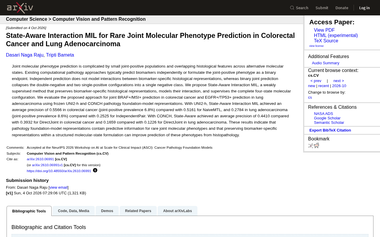
This study addresses the challenge of predicting rare co-occurring molecular phenotypes in computational pathology by proposing State-Aware Interaction MIL. Unlike methods that predict biomarkers independently or collapse multiple negative states into one class, this approach preserves biomarker-specific histological representations, explicitly models their interactions, and applies full supervision over all four molecular configurations. Validated on colorectal cancer (BRAF+/MSI+, 6.8% co-positive rate) and lung adenocarcinoma (EGFR+/TP53+, 8.6% co-positive rate) using frozen UNI2-h and CONCH foundation model representations, it achieves consistent average precision improvements over multiple baselines while demonstrating that pathology foundation models encode meaningful predictive signals even without fine-tuning.
Background: The Challenge of Joint Molecular Phenotype Prediction
Predicting molecular biomarkers from histopathology images has long been a popular direction in computational pathology. But when the research target shifts from single biomarkers to joint molecular phenotypes — where multiple biomarkers are simultaneously positive — the problem becomes considerably harder.
These rare co-occurring phenotypes face two core challenges: first, jointly positive samples are extremely scarce, accounting for only 6.8% of colorectal cancer cases and 8.6% of lung adenocarcinoma cases; second, histological features across different molecular states overlap heavily, making morphology-based differentiation difficult.
Existing computational pathology approaches typically fall into two categories, each with notable limitations. Independent prediction methods predict each biomarker separately but fail to model the interactions between biomarker-specific histological representations. Binary joint prediction methods, on the other hand, collapse double-negative and both single-positive configurations into a single "negative" class, discarding substantial structured information.
BRAF, MSI, EGFR, and TP53 are critical biomarkers in cancer precision medicine. BRAF V600E mutation and microsatellite instability (MSI-H) are commonly co-tested in colorectal cancer because simultaneous positivity suggests a specific oncogenic pathway (the serrated adenoma pathway) and has predictive value for immune checkpoint inhibitor therapy. In lung adenocarcinoma, EGFR mutation is the central criterion for targeted therapy selection (e.g., osimertinib), while TP53 mutation is closely tied to prognosis and resistance mechanisms — their co-mutation carries independent clinical significance. Conventional detection methods, including immunohistochemistry, PCR, and next-generation sequencing (NGS), are costly and resource-intensive in primary care settings. Whether these molecular states can be inferred directly from routine H&E-stained slides is one of the core questions computational pathology seeks to answer, and it is the clinical motivation behind this study.
Methodological Innovation: State-Aware Interaction MIL
The study proposes a weakly supervised method called State-Aware Interaction MIL, whose core idea can be summarized in three points.
First, preserve biomarker-specific histological representations rather than conflating features from different biomarkers. Second, explicitly model the interactions between these representations. Third, apply supervised learning over the complete four-state molecular configuration rather than reducing the problem to binary classification.
The four states refer to the full configuration space: double-negative, two types of single-positive, and the joint double-positive. By retaining this structure, the model can capture fine-grained information that conventional binary methods erase, enabling more targeted identification of those rare jointly positive cases.
The essence of this design is to exploit every histological cue encoded in each molecular state under conditions of extreme class imbalance — rather than crudely collapsing the problem into "positive or negative."
Multiple Instance Learning (MIL) is the core framework in computational pathology for handling weakly supervised problems. Since pathology slide annotations are typically available only at the slide level (i.e., only the patient-level label is known) rather than at the pixel or cell level, MIL treats an entire slide as a "bag" and the image patches cropped from it as "instances." The model learns from bag-level labels without requiring expensive local annotations. Traditional MIL methods typically aggregate all patch features into a slide-level representation via pooling functions (such as max pooling or attention-weighted mean) before classification. The key innovation of State-Aware Interaction MIL lies in maintaining independent representation branches for each biomarker rather than merging all biomarker information into a single aggregation pipeline, then capturing cooperative or antagonistic signals between different molecular states through an explicit interaction mechanism — enabling finer-grained modeling of multi-biomarker joint states under weak supervision.
Experimental Setup and Pathology Foundation Models
The study validated the method in two specific clinical scenarios: BRAF+/MSI+ joint prediction in colorectal cancer and EGFR+/TP53+ joint prediction in lung adenocarcinoma.
For feature representation, the study used two frozen pathology foundation models — UNI2-h and CONCH. "Frozen" here means using representations extracted directly from pre-trained models without fine-tuning, which also serves as an indirect test of the predictive information inherently encoded in pathology foundation model representations.
Average Precision (AP) was chosen as the evaluation metric. In rare phenotype prediction tasks with severe class imbalance, AP is more informative than accuracy, as it better reflects model performance when positive samples are scarce.
Pathology Foundation Models represent a major recent advance in computational pathology — large-scale visual encoders pre-trained on massive pathology image datasets via self-supervised or contrastive learning. UNI2-h and CONCH are representative open pathology foundation models: UNI2-h is pre-trained via self-supervised learning on large-scale pathology images and excels at extracting general tissue morphology features; CONCH employs vision-language contrastive learning to align pathology images with pathology report text, endowing representations with richer semantic content. Using "frozen" representations means the pre-trained models serve as fixed feature extractors with no parameter updates on the downstream task — this substantially reduces the need for annotated data and constitutes a rigorous test of the foundation models' generalization ability: if frozen representations can still support rare joint phenotype prediction, it demonstrates that the pre-training phase has implicitly encoded relevant histological patterns.
Results: Consistent Improvements Across Multiple Baselines
Experimental results show that State-Aware Interaction MIL outperforms corresponding baseline methods across both foundation models and both cancer types.
With UNI2-h representations, the method achieves an average precision of 0.5566 on colorectal cancer, surpassing NaiveMTL's 0.5161; on lung adenocarcinoma it reaches 0.2784, outperforming IndependentPair's 0.2525.
With CONCH representations, average precision on colorectal cancer is 0.4410, an improvement over DirectJoint's 0.3932; on lung adenocarcinoma it is 0.1659, notably higher than DirectJoint's 0.1226.
In absolute terms, the lung adenocarcinoma task is substantially harder than colorectal cancer, with AP scores considerably lower across both foundation models — consistent with the greater complexity of its histological features and the rarity of co-positive cases. Yet regardless of configuration, structured molecular state modeling delivers consistent gains.
Significance and Implications
This study conveys two noteworthy signals.
First, representations from pathology foundation models such as UNI2-h and CONCH do contain information sufficient to predict rare joint molecular phenotypes — even without fine-tuning, frozen representations can support this highly challenging task. This provides further evidence for the practical utility of pathology foundation models.
Second, when modeling rare joint phenotypes, preserving biomarker-specific representations within a structured molecular state framework is more effective than simple independent prediction or binary collapse. This methodological insight carries broader implications for other medical AI tasks involving small samples and multi-label interactions.
For computational pathology and precision medicine, the ability to directly infer rare joint molecular states from routine tissue slides means that valuable auxiliary information can be obtained even when large-scale expensive molecular testing is not feasible. This is precisely the direction that weakly supervised pathology AI continues to explore.
Related articles

Chain of Thought Isn't Just for AI: How Humans Can Use CoT to Improve Thinking Quality
Chain of Thought isn't just a prompt trick for large models. Starting from a Hacker News discussion, this article analyzes why CoT works for human thinking too, breaks down actionable ways to transfer it, and highlights the key differences between human CoT and machine CoT.
Cloudflare Teams Up with OpenAI: Closi…
Cloudflare Teams Up with OpenAI: Closing the Loop from Vulnerability Discovery to Defense with AI
Cloudflare uses OpenAI Daybreak to investigate security vulnerabilities and generate reviewable remediation plans, while developers use Codex to boost coding efficiency and quality.

Orca Multi-Agent Orchestration in Practice: A Guide to Parallel Collaboration with Claude and Codex
Orca is Stable AI's free open-source agent workbench supporting 34 agents like Claude Code and Codex in parallel. Learn its Git worktree isolation, multi-model orchestration, and mobile management.