xvr Technology: How AI Makes Minimally Invasive Surgery Navigation More Precise and Safe
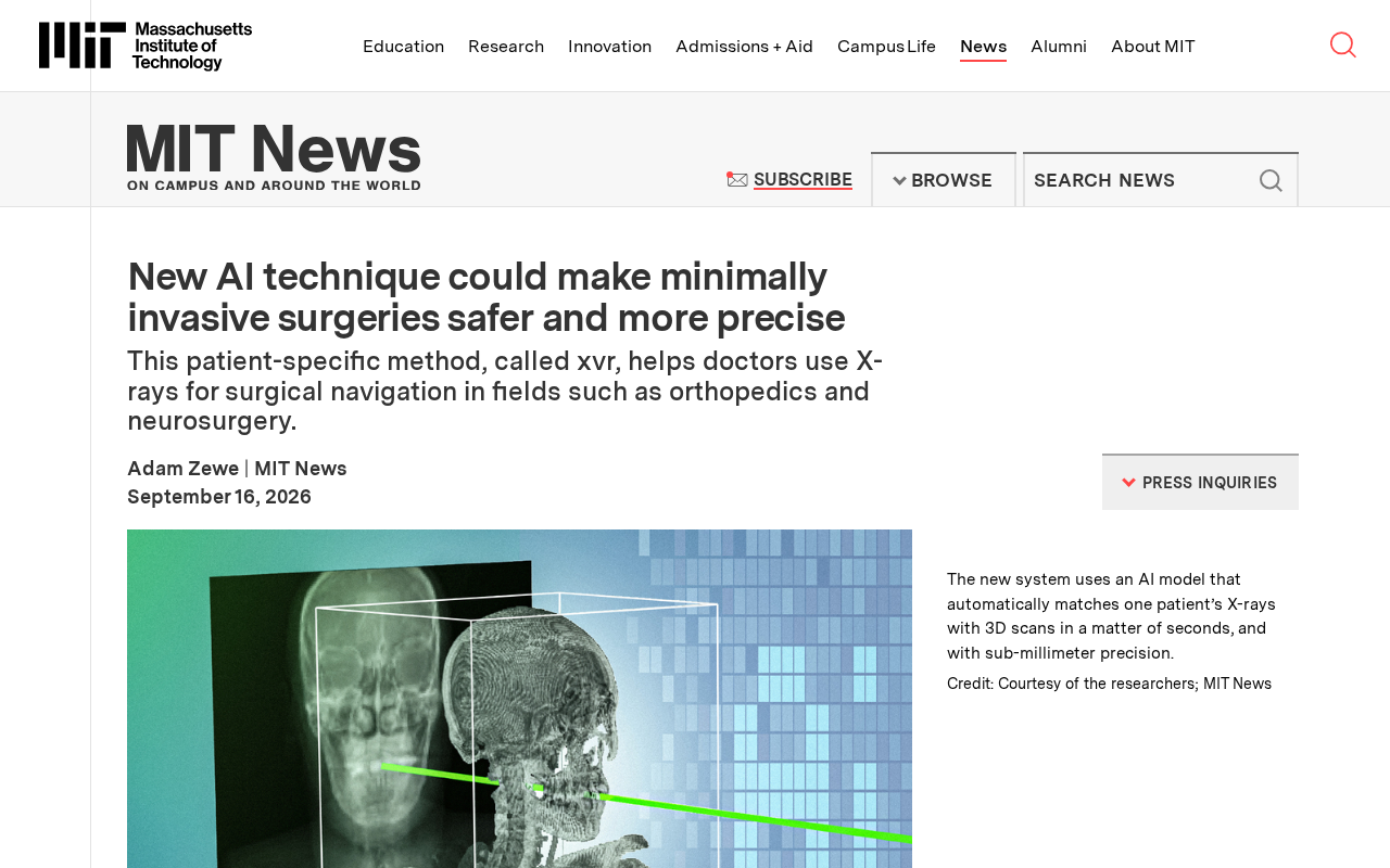
xvr is a patient-specific AI method that precisely aligns intraoperative 2D X-rays with 3D anatomy to improve minimally invasive surgical navigation.
Minimally invasive surgery demands extreme instrument positioning accuracy, and aligning preoperative 3D CT scans with intraoperative 2D X-rays — known as 2D/3D image registration — is a core challenge in surgical navigation. xvr is a patient-specific AI-assisted navigation method that, unlike general-purpose algorithms, optimizes based on each patient's individual anatomy to achieve more precise spatial registration. Targeting high-precision procedures such as spinal screw placement and neurosurgery, xvr aims to reduce repeated intraoperative fluoroscopy, lower radiation exposure, and improve overall surgical safety — reflecting the broader trend of medical AI shifting from universal models toward personalized adaptation.
The Navigation Challenge in Minimally Invasive Surgery
Minimally invasive surgery has become a major trend in modern medicine thanks to its smaller incisions and faster recovery times — but it demands exceptional precision from surgeons. In fields like orthopedics and neurosurgery, surgeons often rely on X-ray imaging to track the real-time position of instruments inside the body. However, the relationship between a 2D X-ray image and a patient's 3D anatomy is far from intuitive. Accurately aligning preoperative 3D CT data with intraoperative 2D X-rays has long been one of the central challenges in surgical navigation.
A new AI technology called xvr is attempting to solve exactly this problem. It offers a patient-specific approach that helps surgeons make better use of X-ray imaging for surgical navigation, making minimally invasive procedures safer and more precise.
The process of aligning preoperative CT scans with intraoperative X-rays is technically known as 2D/3D image registration. The core difficulty lies in the fact that an X-ray is a 2D projection of a 3D structure along a single direction — meaning a single X-ray image could correspond to an infinite number of possible 3D poses. This inherent ambiguity makes the registration problem fundamentally ill-posed. Traditional methods typically rely on manually annotating anatomical landmarks or iteratively adjusting virtual projection angles to match the real X-ray — a process that is time-consuming and highly operator-dependent. In recent years, deep learning-based methods have attempted to directly regress the 6-DoF pose (three translations and three rotations) of instruments or bones from images, but poor generalization across different patients has continued to limit clinical adoption.
What Is xvr?
In simple terms, xvr is an AI-assisted navigation method tailored to each individual patient. Conventional surgical navigation algorithms typically rely on general-purpose models that struggle to fully accommodate the unique anatomical variations of each patient. xvr emphasizes a patient-specific approach — optimizing based on the imaging data of a specific individual — to achieve more accurate spatial registration between X-ray images and 3D anatomical models.
The value of this approach lies in a fundamental mismatch: during surgery, what the surgeon sees is a real-time 2D X-ray, but what they truly need to understand is the 3D position of their instruments. xvr acts as a bridge between the two, precisely linking intraoperative X-ray images with the patient's own 3D model to provide surgeons with a more reliable spatial reference.
The patient-specific approach is typically implemented as follows: before surgery, the patient's own CT data is used to build a personalized 3D model, which is then used to generate a large number of simulated X-ray projections — known as Digitally Reconstructed Radiographs (DRRs) — to serve as training or optimization data. Because the model is derived from that patient's own anatomy, the algorithm does not need to generalize across a population; it only needs to solve for the pose within that patient's anatomical constraints, significantly reducing registration error. This workflow also differs from the paradigm of "train a general model, then run inference directly" — xvr requires a patient-specific preprocessing or fine-tuning step to be completed before each procedure.
Applications in Orthopedics and Neurosurgery
Based on available information, xvr is primarily targeting surgical fields with extremely high precision requirements, including orthopedics and neurosurgery.
In orthopedic procedures — such as spinal pedicle screw placement or fracture reduction and fixation — even minor deviations in instrument positioning can lead to serious consequences. Neurosurgery demands even greater care: the tissues surrounding the brain and spinal cord are highly fragile, and any navigation error can cause irreversible damage. In these contexts, more precise X-ray guidance translates directly to reduced procedural risk and better surgical outcomes.
By providing customized navigation support for each patient, xvr has the potential to reduce the number of repeated intraoperative X-ray acquisitions and exploratory maneuvers. This could not only improve surgical efficiency but also lower the radiation exposure for both patients and surgical staff.
Pedicle screw placement in spinal surgery is one of the most representative scenarios for demanding navigation precision. The pedicle — the bony channel connecting the vertebral body to the vertebral arch — is only a few millimeters in diameter. A screw that deviates outside this channel can penetrate the spinal cord or nerve roots, causing paralysis or severe pain. Existing research shows that even with standard fluoroscopic navigation, pedicle screw misplacement rates remain between 5% and 15%. Reducing this rate is precisely where high-precision X-ray navigation methods deliver their core value. The clinical significance of reducing intraoperative fluoroscopy is equally important: the radiation dose from a complex spinal procedure can be equivalent to hundreds of standard chest X-rays, and the cumulative occupational exposure risk for surgeons over a career is a well-recognized concern in the surgical community.
Technical Significance and Outlook
xvr represents an exploration of the convergence between AI, medical imaging, and surgical navigation. In recent years, AI applications in medical image analysis have expanded steadily — from lesion detection and image segmentation to surgical planning — gradually permeating every stage of the clinical workflow. Surgical navigation, which demands both real-time performance and high precision, is one of the most challenging yet high-value areas for AI deployment.
Notably, the rise of patient-specific methods reflects a broader shift in medical AI: from general-purpose models toward personalized adaptation. Every person's skeletal morphology and tissue structure is different, and algorithms that can optimize for the individual are often better suited to meet the stringent demands of clinical practice than one-size-fits-all solutions.
Of course, there is still a significant journey from technical validation to large-scale clinical adoption. Methods like this require rigorous clinical trials and regulatory approval before they can truly enter the operating room. But it is reasonable to anticipate that as AI-powered navigation technology matures, both the safety margins and the precision ceiling of minimally invasive surgery will continue to expand — and ultimately, it is patients who will benefit most.
Summary
xvr demonstrates the potential of AI to enhance surgical safety. By focusing on patient-specific X-ray navigation, it addresses the high-precision demands of orthopedics and neurosurgery, providing surgeons with more reliable spatial references during minimally invasive procedures. While publicly available information remains limited, this direction is undoubtedly worth continued attention from both the medical and AI communities.
Related articles

Gemini Enterprise Free Trial Review: Key Features and Usage Considerations
Google's Gemini Enterprise Business tier offers a 30-day free trial with multimodal AI including chat, image, and video generation. We break down features, usage, and the compliance risks behind "unlimited free" claims.

Building a Reasoning Model From Scratch: Motivations and Setup from Sebastian's Lecture 1
Sebastian Raschka's new course Build A Reasoning Model From Scratch: tracing the LLM-to-reasoning-model evolution, the case for from-scratch learning, and UV-based PyTorch environment setup.

AI Agents Keep Acting on Stale Decisions? The Problem Isn't Retrieval — It's Memory Conflict
A developer's hard lesson: AI coding agents acting on stale decisions isn't a retrieval problem — it's memory conflict. How a 200-line freshness scorer failed, and why "newest write wins" works.