94 Deep Learning Pipelines Expose the Medical CV Deployment Crisis: Accuracy Is There, But Where's the Deployment?
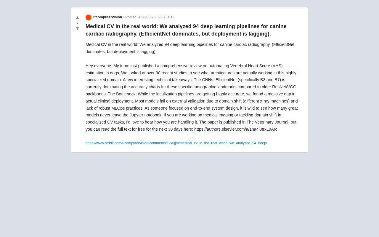
94 deep learning pipelines show medical CV accuracy is solved, but deployment and generalization are not.
A systematic review of 94 deep learning pipelines for canine cardiac radiography reveals EfficientNet as the accuracy leader, but exposes a critical deployment gap. Domain shift across imaging devices and the absence of MLOps practices prevent most models from leaving the lab. The study highlights that bridging this gap requires multi-source training data, domain adaptation techniques, and robust engineering infrastructure.
In the field of medical imaging AI, we often hear good news about model accuracy breaking new records, yet rarely does anyone seriously ask: how many of these models have actually made it out of Jupyter Notebooks and into clinical practice? Recently, a research team published a review in The Veterinary Journal, systematically analyzing 94 deep learning pipelines for canine cardiac radiographic imaging, focusing on the highly specialized task of automated Vertebral Heart Score (VHS) estimation. The conclusions are both exciting and sobering: EfficientNet leads the pack in accuracy, but actual clinical deployment is severely lagging behind.
What Is VHS: AI's Entry Point into Veterinary Cardiac Imaging
Vertebral Heart Score (VHS) is the core quantitative metric for assessing canine heart size in veterinary cardiology. First proposed by Buchanan and Bücheler in 1995, it remains the most widely used quantitative method for evaluating heart size in veterinary radiology. Veterinarians measure the long and short axes of the heart on chest X-rays, normalizing them against vertebral body lengths to determine whether conditions like cardiac enlargement are present.
The specific procedure involves: on a standard right lateral recumbent chest X-ray, measuring the distance from the carina along the cardiac long axis to the apex, then measuring the maximum cardiac width perpendicular to the long axis. These two lengths are projected onto the spine starting from the fourth thoracic vertebra (T4), read in vertebral units, and summed. Normal canine VHS typically falls within 9.7±0.5 vertebral units, though significant breed-specific variation exists—for example, Dachshunds have higher normal values while Whippets trend lower. This inter-breed variation itself increases the technical difficulty of automated assessment and places higher demands on model generalization.
This task is essentially a radiological landmark localization + geometric measurement problem: it requires precise identification of specific anatomical landmarks on the cardiac silhouette and vertebral positions. Landmark localization is a classic computer vision task, with two major paradigms: heatmap regression methods—generating a probability heatmap for each keypoint where the peak position represents the predicted coordinates; and direct coordinate regression methods—where the network directly outputs coordinate values. In medical imaging, heatmap methods are more commonly used due to their superior spatial information preservation, though they correspondingly require higher-resolution inputs. VHS measurement requires simultaneously localizing key anatomical points on the cardiac silhouette and boundary points of spinal vertebrae, involving multiple spatially dispersed targets—making it more challenging than single-region keypoint detection (such as facial landmarks).
Traditionally, this measurement is highly dependent on veterinary expertise, subjective, time-consuming, and difficult to standardize. This is precisely why it's a natural application scenario for computer vision automation—it has clearly defined quantitative objectives and genuine clinical demand.
Why EfficientNet Dominates Medical Imaging Accuracy Leaderboards
After reviewing over 90 recent studies, the research team found that in backbone selection, EfficientNet (particularly the B3 and B7 variants) significantly outperformed earlier ResNet and VGG architectures in accuracy.
This result is unsurprising. EfficientNet was proposed in 2019 by Mingxing Tan and Quoc V. Le from Google Brain, with the core idea of using compound scaling to systematically balance network depth, width, and input resolution, achieving superior feature extraction capability at equivalent parameter counts. Traditional approaches typically scale only a single dimension of the network—for instance, deepening the network (like ResNet scaling from 18 to 152 layers), widening channels, or increasing input resolution. EfficientNet's compound scaling coordinately expands all three dimensions by a fixed ratio, mathematically expressed as: depth d=α^φ, width w=β^φ, resolution r=γ^φ, where α, β, γ are determined through grid search on the baseline model (B0), and φ is the user-controlled scaling coefficient. B0 through B7 represent configurations from φ=0 to φ=7; B7 reaches an input resolution of 600×600 with approximately 66 million parameters, while B3 has only about 12 million parameters at 300×300 resolution.
For tasks like VHS that require capturing fine-grained anatomical landmarks, precise localization at high resolution is particularly critical—B7's greater capacity and resolution adaptability show clear advantages here, while B3 offers a more practical trade-off between accuracy and computational cost. This design philosophy enabled EfficientNet to achieve state-of-the-art accuracy on ImageNet with far fewer parameters and FLOPs than comparable models, and its pretrained weights perform exceptionally well when transfer-learned to small-dataset domains like medical imaging.
By comparison, classic architectures like VGG suffer from parameter redundancy and lower feature representation efficiency. ResNet, while solving deep training challenges through residual connections, has been surpassed by newer architectures in localization accuracy within this specific subdomain. This also reflects a broader trend: even in highly specialized medical subfields, the iterative gains from mainstream architecture advances continue to pay dividends.
The Real Bottleneck: The Deployment Gap Far Exceeds the Accuracy Gap
However, the most valuable insight from this review isn't about "which model is most accurate"—it's about exposing a reality long overlooked by the industry: localization pipelines are becoming increasingly precise, but a massive gap exists in actual clinical deployment.
The research team pointed out that most models fail during external validation, primarily for two reasons:
Domain Shift: The Fatal Challenge of Cross-Device Generalization
Different X-ray machines exhibit systematic differences in imaging parameters, contrast, resolution, and noise characteristics. A model trained to near-perfection on data from Hospital A's equipment may suffer drastic accuracy drops when deployed on Hospital B's machines. This is a textbook example of insufficient generalization in medical imaging AI—models may be learning the imaging "fingerprint" of specific devices rather than truly robust anatomical features.
From a probabilistic perspective, the conditional probability P(Y|X) learned by a model on the source domain (training data distribution P_source) may no longer hold when the input data distribution shifts to P_target. In medical imaging scenarios, sources of domain shift are extremely diverse: different X-ray equipment brands (e.g., Fujifilm vs. Carestream) use different detector technologies and image post-processing algorithms; differences in tube voltage (kVp) and tube current (mAs) directly affect image contrast and noise levels; systematic imaging characteristic differences exist between digital radiography (DR) and computed radiography (CR); even the same device under different calibration states may produce distribution drift. Due to their powerful feature fitting capabilities, deep learning models are highly prone to encoding these device-specific low-level texture patterns as "shortcut features" into their decisions, resulting in severely compromised cross-device generalization.
Absent MLOps Practices: Models Stuck in the Lab
The deeper issue lies at the engineering level. The vast majority of studies stop at validation metrics reported in papers, lacking robust MLOps practices: no continuous data distribution monitoring, no retraining pipelines for new devices, no reliable version management and deployment pipelines. In the original authors' words, "It's truly disheartening to see so many excellent models that never left Jupyter Notebook."
MLOps (Machine Learning Operations) is a practice framework that brings DevOps principles to machine learning lifecycle management, encompassing data version control, experiment tracking, model registries, automated training pipelines, continuous integration/continuous deployment (CI/CD), model monitoring and alerting, and more. In medical AI, the importance of MLOps is particularly pronounced: first, medical data distributions continuously drift due to equipment upgrades, acquisition protocol changes, and patient population shifts, requiring timely degradation detection through data distribution monitoring (e.g., using Population Stability Index or KL divergence); second, regulatory frameworks like the FDA and EU MDR impose strict traceability requirements on AI medical devices, demanding complete audit trails for every model update; additionally, retraining pipelines must integrate new data without violating data privacy regulations (such as HIPAA and GDPR). Common MLOps toolchains include MLflow for experiment tracking, DVC for data version control, Kubeflow or Airflow for pipeline orchestration, and Prometheus+Grafana for production monitoring. Without this infrastructure, even the most accurate models cannot safely and reliably serve clinical practice.
From "It Runs" to "It Ships": Bridging the Medical AI Deployment Gap
The significance of this study extends beyond veterinary imaging—it reflects a common dilemma across the entire medical computer vision field and broader AI applications. Academia's incentive structures naturally favor "leaderboard climbing"—pursuing higher accuracy numbers on fixed datasets—while the robustness, cross-device generalization, and long-term maintainability required for clinical deployment often receive insufficient attention and investment.
For practitioners focused on end-to-end system design, this raises several directions worth deep consideration:
- Data level: Incorporate multi-device, multi-source data during training to proactively address domain shift, rather than discovering problems only at deployment.
- Technical level: Explore domain adaptation, data augmentation, and domain generalization methods to improve model adaptability to unseen devices. Domain adaptation assumes access to target domain data (typically unlabeled), aligning source and target domain feature distributions through adversarial training (such as gradient reversal layers in DANN), Maximum Mean Discrepancy (MMD) minimization, or optimal transport-based methods. Domain generalization is even more stringent, requiring models to generalize without ever seeing target domain data; common approaches include meta-learning, invariant feature learning based on causal inference, and multi-source domain augmentation (e.g., MixStyle, cross-domain style transfer). In medical imaging, frequency-domain data augmentation—simulating device differences by swapping low-frequency components between images from different domains—has shown promising results across multiple benchmarks. Additionally, Test-Time Adaptation (TTA), as a lightweight compromise, allows models to perform online updates of batch normalization parameters using small amounts of target domain data during inference—another practical technique worth attention.
- Engineering level: Transform MLOps from an "optional nice-to-have" into a "mandatory requirement," establishing a complete closed loop from data collection through model monitoring to retraining.
Conclusion
This review published in The Veterinary Journal sounds an alarm for the industry through its analysis of 94 real-world pipelines: the ceiling of model accuracy keeps rising, but the floor of deployment capability has yet to be firmly established. EfficientNet's dominance demonstrates that architecture selection is maturing, while the widespread failure of external validation reminds us that the next breakthrough in medical AI may not lie in fancier networks, but in more solid deployment engineering and generalization capabilities. For any team battling domain shift in specialized CV tasks, this offers a valuable portrait of reality worth studying.
Related articles

AI Agent Cost Optimization in Practice: Engineering Wisdom That Saved $1 Million in One Hour
Databricks eliminated $1M/year in wasted AI Agent spend in just one hour. Learn the root causes of Agent cost overruns and key strategies like model tiering, context pruning, and caching.

How the FDA Is Building an AI-Ready Data Foundation on Databricks
Explore how the FDA leverages Databricks for Government to build a unified Lakehouse architecture and AI-ready data foundation while meeting federal security and compliance standards.

The Power of Security Collaboration: Why Vulnerability Discovery Cannot Do Without Human Intelligence
Explore how security collaboration outperforms tool dependency, the value of vulnerability stories, cross-team knowledge sharing practices, and building stronger defenses by investing in people and collaboration.