Deep Learning on Brain DICOM Datasets: A Guide to Choosing Between 2D and 3D Approaches
Practical guide to 2D vs 3D deep learning approaches for brain DICOM datasets with ADNI preprocessing
A comprehensive guide addressing the core methodological dilemma in brain medical imaging deep learning: choosing between 2D slice-based, 2.5D multi-slice, and full 3D CNN approaches. Covers DICOM format specifics, ADNI dataset characteristics, and standard preprocessing workflows including resampling, MNI registration, skull stripping, and intensity normalization.
Starting from Scratch: The Deep Learning Dilemma in Brain Medical Imaging
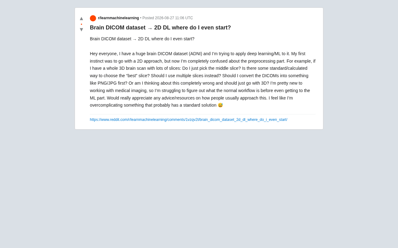
Recently, in Reddit's machine learning community, a novice researcher raised a very typical question: armed with a massive brain DICOM dataset (ADNI, the Alzheimer's Disease Neuroimaging Initiative dataset), wanting to apply deep learning techniques, yet feeling lost at the most fundamental preprocessing stage.
Their confusion centered on several core questions: faced with a 3D brain scan containing numerous slices, should they just take the middle slice? Is there a standard method to select the "best" slice? Should they use multiple slices instead? Do they need to convert DICOM to PNG/JPG format first? Or should they go directly with a 3D approach from the start?
This seemingly simple question actually touches on the most critical methodological choices in medical imaging deep learning. As the questioner self-deprecatingly remarked: "I feel like I'm overcomplicating what should have a straightforward answer." In fact, this is precisely the crossroads every person entering the medical imaging AI field will encounter.
The Peculiarities of DICOM Format and ADNI Dataset
What is DICOM Format
DICOM (Digital Imaging and Communications in Medicine) is the international standard format for medical imaging. Unlike ordinary JPG/PNG images, DICOM files contain not only pixel data but also extensive metadata, such as scan parameters, patient information, spatial coordinates, pixel spacing, slice thickness, etc. This information is crucial for correct interpretation of medical images.
The DICOM standard was jointly developed by the American College of Radiology (ACR) and the National Electrical Manufacturers Association (NEMA), and has become the global standard for medical institutions since the release of version 3.0 in 1993. A DICOM file internally consists of a series of Data Elements, each identified by a Tag, for example (0028,0010) represents the number of image rows, and (0028,0030) represents pixel spacing. DICOM also defines network communication protocols (such as C-STORE, C-FIND), enabling devices from different manufacturers to exchange images. In deep learning scenarios, understanding DICOM's Transfer Syntax is particularly important, as it determines the compression method and byte order of pixel data, directly affecting the correctness of data reading.
The Research Value of ADNI Dataset
ADNI is one of the world's most renowned Alzheimer's disease research datasets, containing extensive structural MRI, functional MRI, and PET scan data. A notable characteristic of this type of data is that pathological information is often distributed throughout the entire three-dimensional space, rather than concentrated in a single slice. This characteristic directly influences the choice of subsequent deep learning methods.
Since its launch in 2004, the ADNI project has gone through four phases (ADNI-1, ADNI-GO, ADNI-2, ADNI-3), enrolling over 2000 subjects in total, covering three groups: cognitively normal elderly, mild cognitive impairment (MCI), and Alzheimer's disease patients. The structural MRI of this dataset typically uses T1-weighted sequences, with voxel resolution around 1mm×1mm×1mm, and a single 3D scan contains approximately 170-200 sagittal slices. ADNI's unique value lies in its longitudinal tracking design—the same subject undergoes follow-up scans every 6-12 months, enabling researchers to analyze the trajectory of brain structure degradation over time, which is extremely critical for training deep learning models that predict disease progression.
2D or 3D? The Core Methodological Choice
The Trap of Simply Taking the Middle Slice
For beginners, the most intuitive "take the middle slice" approach requires caution. While the middle slice does anatomically contain more brain tissue structures, for diagnostic tasks like Alzheimer's disease, key information about pathology (such as hippocampal atrophy, ventricular enlargement) may be distributed in specific slices that are not in the middle position. Simply fixing on a certain slice will likely lose critical diagnostic information.
Reasonable Implementation of 2D Methods
If a 2D approach must be adopted (usually due to computational resource constraints or data volume considerations), there are several more mature industry practices:
- Multi-slice sampling: Rather than limiting to a single slice, extract multiple slices from each scan (for example, consecutive slices around the region of interest), treating them as independent samples or combined inputs.
- 2.5D method: Stack adjacent multiple slices into an input similar to RGB three channels, allowing the 2D convolutional network to indirectly obtain partial spatial context information. This is a compromise between efficiency and performance. The core idea of the 2.5D method is to provide the network with a certain degree of three-dimensional context without introducing the high computational cost of 3D convolution. The most common implementation is to take the target slice plus one adjacent slice before and after, placing the three slices into the three RGB channels respectively. This way, pre-trained 2D networks (such as ResNet trained on ImageNet) can be directly reused, while the network can implicitly learn inter-slice continuity from adjacent slices. Another variant is the tri-planar method, which extracts slices from axial, coronal, and sagittal directions at the same voxel position, fusing features from three perspectives for classification. This method has shown performance close to full 3D approaches in both brain tumor segmentation and Alzheimer's disease classification tasks.
- Projection methods: Such as Maximum Intensity Projection (MIP), compressing 3D volumes into 2D images.
The Advantages and Costs of 3D CNN
For spatially rich data like ADNI, 3D Convolutional Neural Networks (3D CNN) can directly learn spatial relationships between voxels, typically performing better on diagnostic tasks. But the cost is also obvious: computational resource requirements increase dramatically, training time is longer, and memory demands are higher. For beginners, the debugging difficulty of 3D approaches is also relatively greater.
The core of 3D CNN is extending 2D convolution kernels to three dimensions (such as 3×3×3), directly sliding on volumetric data to extract features. Representative architectures include 3D versions of ResNet and VGG, as well as V-Net and nnU-Net designed specifically for medical imaging. Taking nnU-Net as an example, it achieves top performance on multiple medical imaging segmentation benchmarks through automated configuration (including network topology, batch size, patch size, etc.), and is considered a strong baseline for medical image segmentation. The memory consumption of 3D CNN is typically tens of times that of the 2D version—a 256×256×256 3D input can easily occupy tens of GB of memory after a few convolutional layers. Therefore, in practice, patch-based training strategies are often adopted, inputting only a local block of the volume each time (such as 64×64×64), covering the complete volume through a sliding window approach.
Standard Preprocessing Workflow for Medical Imaging
Should You Convert to PNG/JPG
Regarding the question "do I need to convert to PNG/JPG first," the answer is usually not recommended for direct conversion. The reasons are:
- DICOM pixel values are often 16-bit or even higher precision, while PNG/JPG typically only have 8 bits, causing information loss during direct conversion.
- Conversion loses important metadata (such as pixel spacing, slice position).
A more standard practice is to use specialized libraries (such as pydicom, SimpleITK, or nibabel in Python) to directly read DICOM/NIfTI data, processing while retaining full precision, and finally converting to NumPy arrays for use by deep learning frameworks.
Typical Preprocessing Steps
A standard medical imaging preprocessing workflow typically includes:
- Format reading: Use
pydicomornibabelto read raw data. - Resampling: Unify different scans to the same voxel spacing, eliminating device differences.
- Registration: Align all brain scans to a standard space (such as MNI template), which is particularly important for brain research. MNI space (Montreal Neurological Institute space) is the most widely used standard coordinate system in neuroimaging research, built based on the average of hundreds of normal human brain MRIs, defining a unified anatomical reference framework. The process of registering individual brain scans to MNI space typically includes: first performing linear registration through affine transformation (12 degrees of freedom, covering translation, rotation, scaling, and shearing) to eliminate differences in head size and orientation; then performing fine alignment of local anatomical structures through non-linear registration (such as the SyN algorithm). This step is crucial for Alzheimer's disease research because only in a unified space can volume changes in key structures like the hippocampus be meaningfully compared across different subjects. Commonly used registration tools include ANTs (Advanced Normalization Tools), FSL's FLIRT/FNIRT, and the recon-all pipeline in FreeSurfer.
- Skull Stripping: Remove parts outside brain tissue to reduce interference. Skull stripping is an indispensable step in brain imaging preprocessing, aimed at removing non-brain tissue such as skull, scalp, and eyeballs from MRI images. Without this processing, deep learning models may learn features unrelated to diagnosis—for example, differences in skull thickness or scalp fat across individuals, which may be statistically correlated with age and thus become confounding factors. Classic skull stripping algorithms include FSL's BET (Brain Extraction Tool, based on deformable models), FreeSurfer's watershed algorithm, and recent deep learning-based methods such as SynthStrip and HD-BET. Deep learning methods are superior to traditional methods in both robustness and speed, especially when processing pathological brains (such as significant deformations caused by tumors).
- Intensity Normalization: Since MRI signal intensity has no absolute standard, normalization is an essential step. Unlike CT imaging which has clear Hounsfield units, MRI signal intensity is in arbitrary units, and even the same device scanning the same person at different times may yield different absolute intensity values. Common normalization methods include Z-score standardization (subtract mean and divide by standard deviation), percentile clipping (such as linear scaling to [0,1] after removing extreme values in the top and bottom 1%), and more advanced histogram matching (aligning the intensity distribution of all samples to a reference sample).
- Cropping and scaling: Unify dimensions to fit network input.
Practical Advice for Medical Imaging Deep Learning Beginners
Leverage Mature Open Source Tools
The medical imaging field has abundant mature open source tools and preprocessing pipelines. For example, MONAI (Medical Open Network for AI) is a framework designed specifically for medical imaging deep learning, with built-in complete toolchains from data loading, augmentation to model training, greatly lowering the entry barrier. MONAI was jointly initiated by NVIDIA and King's College London, built on PyTorch. Its core components include: the transforms module (providing over 100 medical imaging-specific data transformations, such as random elastic deformation, random affine transformation, intensity perturbation, etc.); the networks module (with built-in 3D versions of classic and cutting-edge architectures such as UNet, SegResNet, SwinUNETR); and the data module (supporting intelligent cached loading of multiple formats including DICOM, NIfTI, PNG, significantly reducing I/O bottlenecks). MONAI also provides engineering support such as automatic mixed precision training and distributed training, allowing researchers to focus on scientific questions rather than engineering details. MONAI Label, launched in 2023, further supports interactive annotation workflows and can seamlessly integrate with visualization tools like 3D Slicer.
FSL, FreeSurfer, and other professional neuroimaging software also provide mature brain processing workflows. FSL (FMRIB Software Library) is a comprehensive neuroimaging analysis toolkit developed by Oxford University, covering complete functions from basic preprocessing to advanced statistical analysis. FreeSurfer is renowned for its precise cortical surface reconstruction and automatic anatomical segmentation capabilities. Its recon-all pipeline can automatically complete a full suite of analyses from skull stripping to cortical thickness measurement, although processing a single subject may take 6-12 hours.
A Progressive Learning Strategy
For beginners, a progressive route is recommended: first quickly run through the entire process with a 2.5D or multi-slice 2D approach to build intuition about the data and task; after establishing a baseline, then try the 3D approach for performance improvement. This avoids being overwhelmed by complex 3D engineering problems from the start, while gradually approaching the optimal solution.
It's worth mentioning that performing Alzheimer's disease classification on ADNI data is an extensively researched topic, with abundant benchmark results available in the literature. Beginners can first reproduce published paper results to verify whether their preprocessing pipeline and training configuration are correct, then innovate on that basis. Additionally, special attention must be paid to data leakage issues—since ADNI contains multiple longitudinal scans of the same subjects, when splitting training and test sets, division must be by subject rather than by scan, otherwise the model may just "memorize" specific individuals' brain features rather than learning general disease patterns.
Conclusion
This questioner's confusion actually reflects a common challenge in cross-disciplinary research—medical imaging requires both deep learning technology and some knowledge of medical imaging. The good news is that this field already has quite mature methodologies and tool ecosystems. The key is not to rush, but to first understand the characteristics of the data itself, then choose an appropriate technical path. As community experience shows: rather than obsessing over micro-decisions like "which slice to take," it's better to plan the entire project from an overall workflow perspective.
Related articles

Enterprise-Level Agent Development: Six Core Competencies for Interview Success
Deep dive into core interview topics for enterprise-level Agent development, covering streaming interruption handling, high-concurrency architecture, multi-tenant isolation, observability, and other engineering challenges to help AI large model job seekers build complete capability systems.

MIT Proposes CW-Net: Making Autonomous Driving AI Decision-Making Explainable and Predictable
MIT researchers propose CW-Net concept warning network, transforming autonomous driving AI's black-box decisions into human-understandable concepts, enabling error prediction and human-machine collaboration. This article analyzes its working principles and practical implications for regulatory compliance, safety redundancy, and public trust.

AI Fatigue: Why Do Learners Feel More Lost as AI Gets Stronger?
As AI crushes top human competitors in math proofs and programming contests, how should tech learners cope with AI fatigue and career anxiety? This article analyzes the nature of capability squeeze and provides a rational framework for addressing AI anxiety.