SlideRuler: Calibrating Scanner Bias in Pathology Foundation Models with a Single Slide

SlideRuler corrects scanner-induced feature drift in pathology AI using a single slide as its own calibration reference.
Different scanners introduce systematic imaging differences that cause pathology foundation model embeddings to drift across devices, hindering cross-institutional deployment. The arXiv paper SlideRuler learns a transfer map from paired rescans to capture device-specific feature shift patterns, enabling single-slide calibration at inference without modifying the frozen model. Validated on two encoders and five SCORPION scanners, it reduces average target-to-source feature distance by 16.3%–38.5%, while a source-anchored variant cuts source feature displacement by 47.7%–83.6%. Requiring no external reference samples, SlideRuler offers a practical, lightweight path to consistent pathology AI deployment across heterogeneous imaging environments.
Scanner Variability: The Hidden Barrier to Pathology AI Deployment
Pathology Foundation Models are becoming the core engine of digital pathology analysis, yet a long-underestimated problem continues to hinder their widespread deployment — scanner variability. When the same tissue block is imaged on different scanner models, the model generates different feature representations (embeddings). This device-induced shift means a model trained or validated on Scanner A may behave inconsistently when switched to Scanner B.
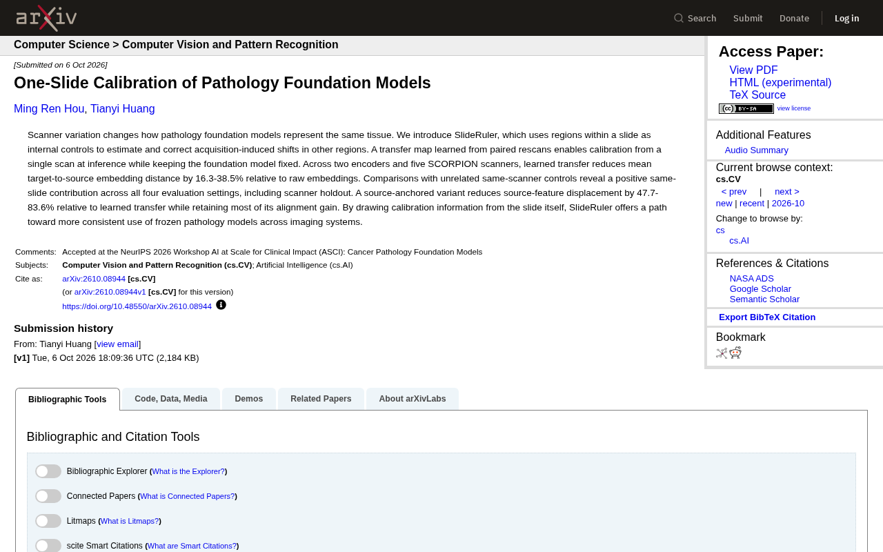
The SlideRuler method, proposed in the recent arXiv paper "One-Slide Calibration of Pathology Foundation Models", directly addresses this pain point. Its core idea is elegantly simple: rather than modifying the already frozen foundation model, it uses regions within the slide itself as "internal references" to estimate and correct the feature drift introduced during acquisition.
Pathology foundation models are typically pre-trained in a self-supervised manner on massive whole slide images (WSIs) to learn universal tissue morphology representations, allowing downstream tasks (such as cancer grading and gene mutation prediction) to benefit from lightweight fine-tuning. However, digital pathology scanners differ in optical design, color calibration, objective magnification, and image compression algorithms, causing systematic deviations in hue, contrast, and even texture for the same tissue across different devices (e.g., Aperio, Hamamatsu, Leica, 3DHISTECH). These deviations may be subtle at the pixel level, yet they are sufficient to cause deep models to extract vastly different high-dimensional feature vectors, ultimately affecting clustering, retrieval, and classification results. Studies have shown that scanner origin can be predicted with high accuracy from embeddings alone, confirming that device fingerprints are deeply encoded by these models. This is precisely why data augmentation alone cannot fully eliminate scanner shift — augmentation only covers known transformation spaces, while real device differences are often far more complex and difficult to enumerate in advance.
The Core Mechanism of SlideRuler
The key to SlideRuler lies in the transfer map. Researchers first learn a transfer map using paired rescans, and then at inference time, only a single scan is needed for calibration — all while keeping the foundation model parameters frozen. The practical value of this design is significant: hospitals do not need to retrain or fine-tune large foundation models for each device; a single calibration pass yields more consistent representations.
In other words, SlideRuler uses the slide itself as a calibration ruler (hence the name). Different regions within a slide share natural internal correlations, and by using these regions as references, the model can infer and compensate for systematic shifts introduced by the scanning device.
Why "Intra-Slide References"?
Conventional cross-device calibration typically requires external reference samples or large amounts of paired data — costly and difficult to generalize. SlideRuler's approach of extracting calibration information from within the slide itself reduces dependence on external references, making single-slide calibration feasible and far easier to integrate into real clinical workflows.
Methodologically, the transfer map is a form of feature space alignment, related to techniques such as linear transform correction in Domain Adaptation and Optimal Transport. Its core assumption is that the feature shift from a source scanner to a target scanner approximates some structured global transformation that can be learned from paired samples. SlideRuler's innovation lies in internalizing the search for paired samples within a single slide — different regions of the same slide share local biological continuity and can serve as soft references for one another, enabling the estimation and application of a correction transform at inference time without any additional scans. This stands in sharp contrast to traditional calibration approaches that rely on external quality control slides (e.g., staining standards) or require the same sample to be scanned repeatedly on multiple devices, dramatically lowering the barrier to clinical deployment.
Experimental Results: Significant Reduction in Feature Distance
The paper validates SlideRuler on two encoders and five SCORPION scanners. Results show that the learned transfer map reduces the average target-to-source feature distance by 16.3% to 38.5% compared to raw embeddings.
The ablation study design is particularly noteworthy. The researchers compare intra-slide references against unrelated same-scanner references and find that "intra-slide contribution" yields positive effects across all four evaluation settings (including scanner holdout). This demonstrates that the calibration gain genuinely stems from the structural information within the slide, rather than incidental scanner-level correlations.
Additionally, the paper introduces a source-anchored variant. Compared to pure learned transfer, this variant further reduces the source feature displacement by 47.7% to 83.6%, while retaining the majority of alignment gains. This means that in scenarios where it is important to keep source features as stable as possible, the source-anchored approach provides additional flexibility.
The SCORPION scanners used in the experiments are a multi-scanner system designed for high-throughput clinical pathology, capable of running multiple units of the same model on a unified platform — meaning subtle inter-unit optical differences still exist even among identical models. Choosing this platform for validation controls for the scanner model variable to focus on individual unit differences, but also means the experiments have not yet covered more extreme cross-brand or cross-imaging-modality scenarios (e.g., brightfield vs. fluorescence). Feature distance (embedding distance), typically measured as cosine or Euclidean distance, reflects the relative positions of two sets of feature vectors in high-dimensional space. Reducing the average target-to-source distance by 16% to 38% means that after calibration, representations of the same tissue region across different devices are more consistent — but the translation of this metric into downstream classification accuracy or survival prediction AUC still needs to be quantified through task-specific experiments.
Implications for Digital Pathology
SlideRuler's greatest value lies in charting a path toward "consistent use of frozen models across devices." As pathology foundation models grow ever larger and more expensive to train, enabling stable performance across different imaging systems without repeated retraining is a critical step toward clinical deployment.
From a methodological standpoint, this work points to a more general insight: when facing domain shift, rather than adjusting a massive model, it may be more effective to seek usable internal structures within the data itself for lightweight calibration. This perspective may have broader relevance beyond medical imaging.
Limitations and Open Questions
The current experiments are based on two encoders and five SCORPION scanners, leaving device and model coverage relatively limited. SlideRuler's performance across a wider range of scanner models, tissue types, and real diagnostic tasks (e.g., downstream classification and segmentation performance) still requires further validation. The paper reports improvements at the embedding distance level; the degree to which these translate into gains in clinical diagnostic accuracy remains a direction worth tracking in follow-up research.
Conclusion
SlideRuler achieves foundation model calibration from a single slide, balancing practicality with methodological novelty. It leaves the frozen model untouched, requires no external standards, and extracts calibration signals from the slide itself — providing a lightweight yet effective solution for cross-device consistency in pathology AI. For researchers and institutions looking to deploy foundation models in heterogeneous scanning environments, this is a development worth watching.
Related articles

Apple HomeView Hands-On Review: Is the $300 Smart Home Hub Worth It?
Apple HomeView hands-on: a $300 smart home hub with facial recognition for quick account switching, but no App Store or voice recognition. Is the wall-mounted version worth it? Should you buy the first generation?

Meta Muse Wonder Launch: What It Means for Meta's Return to the Frontier AI Race
Meta releases Muse Wonder and WonderFull, benchmarks reach the frontier, community feedback is polarized. An in-depth look at Meta's return to the frontier AI race, its personal agent strategy, and Zuckerberg's different path.

Apple Home Smart Home Lineup Leaked: LG and Schneider Team Up, UWB Door Lock on Board
Apple Home ecosystem gains new LG and Schneider devices: UWB smart lock with proximity unlock, 2.5K dual-camera doorbell, privacy indoor camera, and AirPods-like seamless pairing.