Understanding Functional Ultrasound Imaging (fUSI) from Scratch: A New Window into Neuroscience

fUSI uses ultrafast ultrasound to track brain blood flow, enabling high-resolution functional brain imaging at low cost.
Functional Ultrasound Imaging (fUSI) is an emerging brain imaging method that uses high-frame-rate plane wave ultrasound and power Doppler to track microvessel blood flow changes as an indirect measure of neural activity, based on neurovascular coupling. Compared to fMRI, fUSI offers lower cost, a smaller footprint, millisecond temporal resolution, and millimeter spatial resolution, with demonstrated potential in whole-brain animal imaging, neonatal monitoring, and BCI research. Building fUSI from scratch means implementing the full pipeline—from raw RF data through beamforming and SVD-based clutter filtering to functional map generation—advancing open-source development. Key challenges include strong skull-induced ultrasound attenuation in adults and the real-time processing demands of high-frame-rate data.
What Is Functional Ultrasound Imaging (fUSI)?
Functional Ultrasound Imaging (fUSI) is a brain imaging technology that has emerged at the intersection of neuroscience and biomedical engineering in recent years. It uses ultrasound waves to track subtle changes in cerebral blood flow, indirectly reflecting neural activity. Compared to traditional functional MRI (fMRI), fUSI offers unique advantages in temporal and spatial resolution, equipment cost, and portability.
A post shared on Hacker News titled "Functional Ultrasound Imaging (fUSI) from scratch" emphasizes understanding and building this technology from the ground up. Although community engagement is still modest (around 10 upvotes and 1 comment), it points to a technology direction worth watching: how to achieve high-quality brain function observation using relatively affordable, accessible hardware.
The Technical Principles Behind fUSI
fUSI is grounded in the phenomenon of neurovascular coupling — when a brain region becomes active, local blood flow increases to meet energy demands. Using high-frame-rate plane wave transmissions combined with Doppler processing, ultrasound imaging can capture changes in red blood cell flow within the brain's microvasculature.
High-Frame-Rate Ultrasound and Power Doppler
Conventional ultrasound imaging has a limited frame rate that makes it difficult to capture rapid hemodynamic changes. The breakthrough in fUSI lies in adopting ultrafast imaging, which can achieve frame rates of thousands of frames per second. Combined with power Doppler processing, the system statistically accumulates blood flow signals, enabling detection of vessels with diameters as small as tens of micrometers.
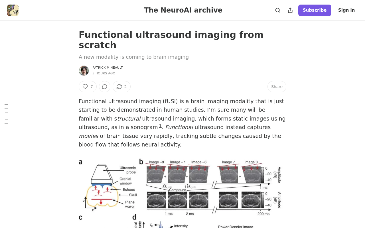
Ultrafast imaging relies on plane wave transmission technology. Traditional focused ultrasound images one scan line at a time, with the frame rate limited by the speed of sound and imaging depth. Plane wave transmission illuminates the entire field of view in a single shot, then uses synthetic aperture algorithms during post-processing to reconstruct the image — boosting frame rates from tens of frames per second to thousands.
Power Doppler works as follows: in a high-frame-rate sequence, echoes from stationary tissue are highly correlated between frames, while moving red blood cells introduce random phase variations. By separating the two using clutter filtering algorithms such as singular value decomposition (SVD), and then integrating the power (squared magnitude) of the residual blood flow signal over time, a power Doppler image is produced that reflects vascular density and blood flow. This pipeline extends traditional Doppler — originally limited to large vessels — down to pre-capillary arterioles with diameters below 50 micrometers.
Comparison with fMRI
fMRI relies on large superconducting magnets, making the equipment expensive, bulky, and subject to strict constraints on participants. fUSI, by contrast, uses a handheld or implantable probe at significantly lower cost, while offering millimeter-level spatial resolution and millisecond-level temporal resolution. This combination of portability and high resolution makes fUSI particularly attractive for animal studies, intraoperative monitoring, and brain-computer interface (BCI) research.
What Does "From Scratch" Mean?
The phrase "from scratch" in the title implies a pedagogical or open-source approach: rather than relying on commercial, closed systems, the idea is to start from raw ultrasound radiofrequency (RF) data and progressively implement the full pipeline — beamforming, Doppler filtering, motion correction, and functional map generation.
For researchers and engineers, understanding the signal processing logic at each step from first principles not only helps reproduce existing results but also leaves room for algorithmic innovation. For example, clutter filtering — how to distinguish tissue motion from blood flow signals — is one of the key technical challenges determining fUSI image quality.
Clutter filtering is arguably the most critical step in the fUSI signal processing chain. Slow displacements of brain tissue caused by breathing, heartbeat, or body movement generate "tissue clutter" in the Doppler signal that is far stronger than the blood flow echoes. Early approaches used high-pass temporal filters (such as wall filters) to separate the two, but their effectiveness was limited by the spectral overlap between tissue motion and blood flow velocities. Modern fUSI systems widely adopt SVD-based spatiotemporal filtering: the ultrasound data matrix (organized as a time series) undergoes SVD decomposition, and the leading singular vectors — which capture spatially coherent tissue signals — are discarded. The residual is attributed to blood flow. This approach far outperforms frequency-domain filtering in suppressing low-frequency interference such as respiratory motion, and requires no explicit estimation of tissue motion trajectories. It is an algorithmic node that deserves close attention when implementing fUSI from scratch.
Applications and Challenges
fUSI has already been validated for whole-brain functional imaging in rodents and is gradually expanding toward clinical applications, including neonatal brain monitoring (leveraging the open fontanelle as an acoustic window) and real-time intraoperative neurosurgical imaging. In the BCI field, fUSI is also considered a promising non-invasive or minimally invasive signal-reading candidate.
However, challenges remain significant: the adult skull strongly attenuates and distorts ultrasound waves, limiting transcranial applications; the massive volume of high-frame-rate data places heavy demands on storage and real-time processing; and the mapping relationship between blood flow signals and neural activity still requires more precise modeling.
Skull-induced ultrasound attenuation and aberration are the core bottleneck preventing fUSI from reaching adult clinical applications. The skull's high acoustic impedance and heterogeneous structure attenuates ultrasound energy by approximately 10–20 dB while introducing severe phase distortion, significantly degrading beamforming accuracy. Researchers are exploring two main approaches: the first is adaptive beamforming, which uses prior CT or MRI data to build an acoustic model of the skull and applies phase compensation at the transmit/receive stage; the second involves developing large-aperture transducer arrays operating at lower frequencies (1–2 MHz) to improve transcranial penetration depth, at the cost of reduced spatial resolution. Neonatal brain monitoring is feasible precisely because the fontanelle provides a natural, skull-free acoustic window, allowing high-frequency probes to achieve image quality approaching that of animal experiments.
Conclusion
As a rapidly evolving imaging technology, fUSI draws on knowledge from ultrasound physics, signal processing, and neuroscience. The value of "from scratch" initiatives like this lies in lowering the technical barrier and fostering an open-source ecosystem, enabling more people to participate in the innovation of brain imaging tools. For practitioners in brain-computer interfaces, medical imaging, or signal processing, this is a direction well worth following closely.
Note: This article was written based on a brief Hacker News post with limited discussion. The technical details provided here are supplementary background drawn from publicly available knowledge in the fUSI field.
Related articles

AI Compute Is the New Oil, But the Market Mechanism Has Yet to Take Shape
Hacker News compares AI compute to the new oil, noting the lack of price discovery, concentrated supply, and no hedging tools. The article analyzes structural issues in compute trading and explores a possible three-tier market of spot, contracts, and derivatives, plus the real impact on AI practitioners' cost management.

Running Full iOS 16.5 Firmware on Android Devices: Technical Demo or Marketing Gimmick?
A Hacker News post claims full iOS 16.5 firmware can run on Android, with 315 upvotes. This analysis argues it is more likely a web UI simulation than a true firmware port, and explores the value and risks of such projects.

Etsy Cracks Down on AI Slop: Addressing Buyer and Seller Anxieties
Etsy introduces stricter rules limiting AI-generated content in listings, addressing buyer confusion over AI products and seller fears of original work being diluted by AI slop.